Measures of efficiency in the emergency department observation units

Claudia Cicchini MD, PhD1, Antonio Simone MD2, Valentina Valeriano MD1, Donatella Livoli MD1, Francesco Rocco Pugliese MD3.
1Emergency Physician,
2Chair of the Emergency Department Observation Unit,
3Medical Director of the Department of Emergency Medicine
Department of Emergency Medicine, Sandro Pertini Hospital, Rome, Italy 

Abstract

Introduction
 
There are nearly 120 million visits to emergency department each year; as the population increases and ages, the growth rate for emergency department visits and the admissions created a surge in demand for inpatient beds.
Current health care reforms moved against deficiencies in access, cost and quality of care. The drastic reduction of hospital beds number and the need for more inpatient capacity brings attention to short-stay admissions and to the alternatives for such many patients at lower cost without adverse effects on care and quality.
 
Methods
 
The observation medicine literature was reviewed using PubMed. This article is a synthesis in support of observation units with their measures of efficiency.
 
Conclusions
 
Observation units increase patient safety and satisfaction while decreasing unnecessary inpatient admissions and providing fiscal performance. Pushing more patient volume through the emergency department observation units diverts patients away from inpatient services and mitigates both emergency department and inpatient crowding. Furthermore maximizing the number of patients seen in the emergency department observation unit enables the emergency department to capture many additional observation payments. Of course patient care comes first and an optimal observation unit is designed to prioritize serving patients and not management or finance concerns.
Creating an observation unit is a dominant strategy for institutions with the volume to justify the fixed costs of operating one.

Key words: observation unit, emergency department, emergency medicine, efficiency

Introduction
 
There are enormous pressures to acute care hospital to increase patient access, safety, quality of care and satisfaction without increasing costs. Many institutions are now managing part of the emergency department (ED) patients as outpatients in short-stay observation units (OU), operated by the ED (1-10). Such patients require either further treatment or diagnostics before being safely discharged and usually stay less than 24 hours.
The use of the OU for those hospitals with the volume of ED visits to justify one, is a win-win initiative with the potential to lower costs and improving patients safety and satisfaction without impacting the quality of care.
A dominant strategy is one that does at least as well as every other strategy in all situations but does strictly better than every other strategy in at least one situation: the OU is a dominant strategy, because every aspect of care in this setting is at least equivalent, if not better, to the alternative of inpatient care.
Understanding the financing of an OU is critical to justify its creation.
The aim of the article is to clarify the rationale for and operating characteristics of these units and to provide support for their clinical and amministrative benefits.

Financial aspects of the EDOU USE

The creation or expansion of observation units is usually compared with alternative competing capital projects, i.e. expanding acute care ED beds or inpatients beds, but the ability for EDOUs to deliver care and provide additional risk stratification through efficient resource use and shorter hospital stay centers on a business model that has proven profitable. EDOUs use algorithm-driven care, allowing for standardized rapid treatment and evaluation within the 24-hour window required for observation stays. (11-13) Thus the first goal of the EDOU is to augment the clinical capacity of the ED and to maximize ED efficiency and profitability (14-15). 
For those who are discharged home from the ED the Lazio Region treats their ED stay as a type of outpatient visit: there is a fixed repayment based on the triage color code, regardless of the diagnosis and the type and amount of the used resources (table 1) (16). Placing a patient in the EDOU generates a new single code combining the ED visit and ED observation: it’s a single rate of 275 euro per patient, calculated as the average value of 0-1 day hospitalization minus 15% (17). If we properly select the patients for EDOU on the basis of either the diagnosis and the triage code, we can generate the advantage of a higher-paying code.
Another financial benefit results from patients discharged home from the EDOU: EDOU avoided an inpatient admission that would have potentially resulted in a loss for the hospital. The Lazio Region payments for several common diagnoses (i.e. congestive heart failure) do not fully cover average inpatient hospital costs. By managing patients who would have created a loss for the hospital as an inpatient, EDOUs create value, and an inpatient bed could be filled by a patient with a more profitable diagnosis related groups (DRG) payment. However hospitals must be careful about shifting too much acute care into EDOU, because any EDOU stay that results in inpatient admission (about 20% of EDOU patients) are only paid by a single DRG that includes ED, EDOU and inpatient care.
The potential costs associated with an EDOU include fixed, variable and opportunity costs.
Fixed costs include the new construction or conversion from existing spaces and the maintenance costs, and above all the staffing. Physician and nurse staff tends to be minimal because patients in observation have been selected because they represent a low acuity population amenable to simple care algorithms with a high likelihood of being discharged home.
Variable costs include the direct resources required to care for each patient, i.e. the costs of charting, housekeeping, linens for bed turnaround.
Opportunity costs are the opportunities for profit lost because of resources diverted to the EDOU. For hospital at full capacity, filling an inpatient bed with a patient who could have been observed creates an opportunity cost because profitable patients (i.e. transfers, elective surgery patients) may be deferred as a result.
 
Table 1 Regional refund by the Lazio Region to the hospital based on triage color code (16).
 
Triage color code
Rate per patient (euro)
Dead on arrival
25.82
Red
1032.91
Yellow
309.87
Green
154.94
White
41.32

Measures of observation care

An essential component of a profitable EDOU is operational efficiency. Maximum efficiency in the EDOU and subsequently profitability require optimizing three main operational variables: the occupancy rate, the duration of observation, the discharge to home rate.       
            Occupancy rate
Because it is impossible to exactly match patient arrivals to departures (i.e. time needed for bed turnover), the maximum occupancy rate will always be less than 100%. However an optimal occupancy rate approaching 100% is obviously beneficial for a dedicated EDOU with fixed resources (number of beds, nursing staff).
The Pertini Hospital has a 10-bed EDOU with an average length stay of 12 hours. Let’s assume a bed turnover time of 30 minutes. The theoretical maximum daily patient bed time is 240 hours (24 hours x 10 beds). Nor accounting for bed turnover or variability in arrivals, the theoretical occupancy rate is 100% (20 patients per day at 12 hours per patient, occupied 240 hours or 100%). Accounting for bed turnover time, but assuming perfect arrival times, a more accurate maximum occupancy rate can be calculated: 12,5 hours per patient (12-hour stay and 30-minutes bed turn around) allows for 19.2 patients per day (24 hours / 12.5 hours per patient x 10 beds), or 1.92 patients per bed per day (19.2 patients per day / 10 beds). Thus 19.2 patients x 12 hours per patient = 230.4 hours of patient bed time, or a 96% occupancy rate (230.5/240)
Thus even if a new patient was always ready to take the spot of a patient leaving the EDOU, the maximum occupancy rate would be 96%. Actually the variability of arrivals to the EDOU will push this maximum even lower.
            Duration of observation
The maximum length of stay should be less than 24 hours because stays longer than 1 day are inefficient use of the EDOU. No additional payment is generated for keeping patients longer. The payment for observation is replaced by a single diagnosis related groups (DRG) payment for when patients are admitted to the inpatient service. Thus to maximize EDOU volume, the maximum number of patients can be cared for in the EDOU if every patient stayed more than 6 hours, but never less, and no more than 24 hours.
            Discharge to home rate
Assuming a maximum occupancy rate near to 90% and an optimal length of stay between 6 and 24 hours for all EDOU patients, the discharge to home rate remains the elusive variable to optimize. Obviously the ideal rate would approach 100%, but attention should be payed to avoid the increasing short-stay inpatient admission as it represents inefficient use of resources. However even in the ideal clinical trial settings around 20% of patients evaluated in the EDOU require admission.
The discharge home rate could be used to determine the industry-wide benchmark, but these data should be taken with caution because they may not reflect the various social, psychiatric and geriatric independency issue that often complicate disposition management in EDOU patients. No organizations published benchmark rates to date, even if in 1995 Brillman et al. have already suggested that units with a discharge to home rate less than 70% should question their guidelines for observation (18).

Conclusions

If sufficient patient volume exists in an institution to justify the expense of an EDOU, opening or enlarging one to maximize both clinical utility and profitability should be strongly considered. A 5% to 10% of ED volume can be expected to be managed in an observation unit. Assuming the average length of stay and the bed turnover calculations previously discussed and given a ratio of 5 patients per nurse, the minimum efficient size of a dedicated EDOU should be 5 beds, which translate into a minimum ED volume of 30,000 to 50,000 annual visits. Administrators of hospitals with ED volumes greater than 30,000 annual visits, but without an observation unit, should assess with the ED clinical staff if this additional resource could add clinical and financial sense for that institution. Of course the expected return on investment must be calculated over several years and compared with competing alternatives such as expanding acute care ED space or inpatient capacity.
The EDOU operational metrics of occupancy rate, length of stay and discharge to home rate are intertwined: changing one of them affects the two others. These variables can be optimized by proper patient selection: well-constructed inclusion and exclusion criteria must be utilized in addition to well-established diagnostic and treatment algorithms.
There are two compelling arguments for observation: improved clinical decision making and increased profitability. Moving patients to an EDOU frees up overcrowded ED resources such as acute care treatment rooms, and provides patients appropriate evaluation, treatment and risk stratification. At the same time EDOU can improve the profitability of a hospital admission on the basis of the payment of the Lazio Region for color code in the ED admission and in the EDOU.
Although efficiency maximization is an important consideration of EDOU management, payment parameters mustn’t dictate clinical management. Focus must always be on providing the right care to the right patient in the right place at the right time.
References
  1. Dickfos M, Ibrahim H, Evans A, Franza R. Cohort study on emergency general surgery patients and an observation unit. S Afr J Surg 2017; 55(3): 66.
  2. Peacock F, Beckley P, Clark C, et al. Recommendations for the evaluation and management of observation services: a consensus white paper: the Society of Cardiovascular Patient Care.Crit Pathw Cardiol. 2014 Dec;13(4):163-98
  3. Pena ME, Kazan VM, Helmreich MN, Mace SE.Care of Respiratory Conditions in an Observation Unit.Emerg Med Clin North Am. 2017 Aug;35(3):625-645
  4. Stewart M, Bledsoe J, Madsen T, et al.Utilization and Safety of a Pulmonary Embolism Treatment Protocol in an Emergency Department Observation Unit.Crit Pathw Cardiol. 2015 Sep;14(3):87-9
  5. Mong R, Arciaga GJ, Tan HH. Use of a 23-hour emergency department observation unit for the management of patients with toxic exposures.Emerg Med J. 2017 Nov;34(11):755-760
  6. Wheatley MA.Additional Conditions Amenable to Observation Care.Emerg Med Clin North Am. 2017 Aug;35(3):701-712
  7. Wheatley MA, Ross MA.Care of Neurologic Conditions in an Observation Unit.Emerg Med Clin North Am. 2017 Aug;35(3):603-623
  8. Ham JJ, Ordonez E, Wilkerson RG. Care of Acute Gastrointestinal Conditions in the Observation Unit.Emerg Med Clin North Am. 2017 Aug;35(3):571-587
  9. Abetz JW, Adams NG, Mitra B. Skin and soft tissue infection management failure in the emergency department observation unit: a systematic review.Emerg Med J. 2018 Jan;35(1):56-61
  10. Simone A, Livoli D, Valeriano V, Pugliese F.R. Short Term Observation: 2008-2015 Activity Report And Data Analysis. It J E M 2017 (2), ePub
  11. Ross MA, Hockenberry JM, Mutter R, Barrett M, Wheatley M, Pitts SR. Protocol-driven emergency department observation units offer savings, shorter stays, and reduced admissions.Health Aff (Millwood). 2013 Dec;32(12):2149-56
  12. Komindr A, Baugh CW, Grossman SA, Bohan JS. Key operational characteristics in emergency department observation units: a comparative study between sites in the United States and Asia.Int J Emerg Med. 2014 Feb 5;7(1):6
  13. Gabele D, Bugais C, Laguna J.Medical Observation Units and Emergency Department Collaboration: Improving Patient Throughput.J Nurs Adm. 2016 Jul-Aug;46(7-8):360-5
  14. Epelde F, Iglesias-Lepine ML, Anarte L.[Economic crisis: cost and effectiveness of short stay hospital units].An Sist Sanit Navar. 2012 Sep-Dec;35(3):469-75
  15. Baugh CW, Venkatesh AK, Hilton JA, Samuel PA, Schuur JD, Bohan JS.Making greater use of dedicated hospital observation units for many short-stay patients could save $3.1 billion a year.Health Aff (Millwood). 2012 Oct;31(10):2314-23
  16. Deliberazione Regione Lazio 9/07/2004 n° 602 allegato 6 Sistema di remunerazione dell’emergenza.
  17. Deliberazione Regione Lazio 23/11/07 n° 946 Introduzione dell’Osservazione Breve Intensiva nel Lazio dal 10 gennaio 2008.
  18. Brillman J, Mathers-Dunbar L, Graff L, Joseph T, Leikin JB, Schultz C, et al. Management of observation units. American College of Emergency Physicians.Ann Emerg Med. 1995 Jun;25(6):823-30


Keywords

Thrombotic Thrombocytopenic Purpura, microangiopathic haemolytic anaemia, thrombotic events, multiorgan damage, plasmapheresis.

Introduction

Thrombotic Thrombocytopenic Purpura (TTP) is an acute syndrome characterized by the presence of microangiopathic haemolytic anaemia and thrombocytopenia.
The first case of TTP was described by Moschowitz in 1924 [1]. Recently, between 1982 and 2001, several authors [2-5] identified in the deficiency of the metalloproteinase ADAMTS13 the primary pathogenic cause. TTP is a rare disease [6-8]. it occurs between 30 and 50 years of age, more frequently in the female sex. In the absence of treatment, mortality exceeds 90%, while it is reduced to 10-20% after adequate plasma plasmapheresis or infusion therapy. However, half the deaths are attributable to complications associated with plasmapheresis and hospitalization (sepsis, haemorrhages, thrombosis, etc.) [9]

Case Report

A 36-year-old italian woman came to our observation in the emergency department for abdominal pain, menometrorrhagia and vomiting. The patient had no significant past medical history. She had no significant clinical changes. She was hemodynamically stable.

A first blood workup showed anaemia with haemoglobin (Hb) 7.7 g/dl, with a low-threshold mean corpuscular volume (MCV). A severe new-onset thrombocytopenia was found (laboratory tests of about 3 months ago were normal) with platelet count of 7000 per cubic millimeter. A laboratory framework of haemolysis was also associated, with indirect hyperbilirubinemia of 3.17 mg/dl and lactate dehydrogenase (LDH) of 1649 IU/L. Creatinine was 1.23 mg/dl with azotemia of 67 mg/dl and a mild hyperamylasemia 117 mg/dl. Coagulation tests were normal.The patient did not show any clinical signs and laboratory tests suggestive of infections, presenting normal number of white blood cells (WBC/mmc) and WBC differential. C-reactive Protein was 4.7 mg/dl with a negative procalcitonin (<0.50 ng/ml). The human chorionic gonadotropin (hCG) blood test was negative. Considering the clinical examinations, a therapy based on methylprednisolone (60 mg with 250 ml physiological saline solution iv), tranexamic acid (500 mg in 500 ml 0,9 % saline iv) was performed. Moreover, a transfusion of blood and platelets was carried out. 
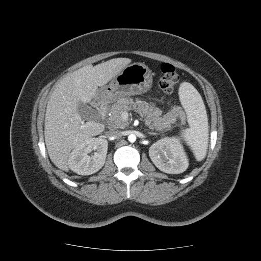
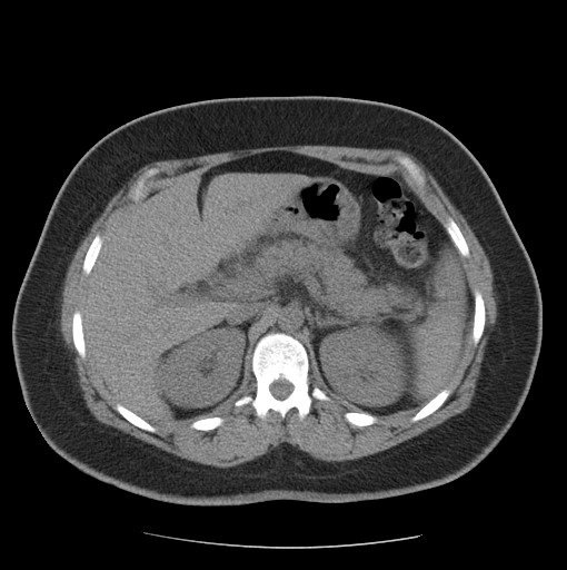
The patient underwent to abdominal and transvaginal ultrasound, which exclude active bleeding and/or pelvic onset diseases, showing a small amount of fluid in the pouch of Douglas. A computed tomography scan (CT) of the abdomen and pelvis was performed with contrast agent and showed “no active bleeding. An increase in pancreatic volume is present with adjacent adenopathies and pervisceral fluid. CT images are suggestive of mild acute pancreatitis and duodenitis.” (Fig. 1, Fig. 2)

Figure 1

Figure 2
 
Moreover, considering the onset of neurological symptoms, such as blurred vision and bilateral intermittent transient hypoacusia, the patient was transferred to a second-level structure and admitted to the complex operative unit of haematology, in the suspect of a microangiopathic disorder.

Discussion

Two different forms of TTP have been classified: Congenital and Acquired forms. Both cases, genetic mutations and immune-mediated mechanisms, are responsable of the ADAMTS13 protein deficiency. The ultralarge multimeter of the von-Willebrand factor (ULvWF) is not appropriately cleaved, causing spontaneous and massive platelet aggregation for intense endothelial stress, especially in the cerebral, cardiac and renal microcirculation. Some predisposing factors are: occult neoplasia or metastatic condition, the intake of drugs, among which the quinidine and ticlopidine [10], and HIV infection [11]. Acquired TTP usually occurs in a previously healthy individual, more commonly in young females as evident in our case report.
The presence of microangiopathic haemolytic anaemia and thrombocytopenia is ample for the diagnosis in the absence of other evident causes [12]. Our patient also presented two of the other three elements of the classical pentade (acute renal failure, neurological changes and fever). These signs are no longer necessary and are associated to advanced organ damage [13]. As in our case, symptomatology can be quite varied, including hemorrhagic events and the most fearful thrombotic manifestations (cardiovascular, renal and neurological). Non specific gastroenterological manifestations, such as fever, abdominal pain, diarrhoea and vomiting are very frequent. Recently McDonald et. [14] described the association with new acute pancreatitis.
Sometimes, the identification of the syndrome can be difficult, being often in overlap conditions with hemolytic-uraemic syndrome (HUS) and autoimmune disorders (antiphospholipid antibody syndrome, Evans syndrome).
Rapid diagnosis must be supported by laboratory tests; the presence of≥1schistocytes on a peripheral blood smearschistocytes is considered suggestive for diagnostic purposes [15, 16]. 
TTP treatment involves the use of plasmapheresis [17] or, in alternative, plasma infusion by evaluating the individual fluid load tolerance [18]. These therapeutic approaches reduce mortality from 80-90% to 10% [9]. The effectiveness of plasma exchange therapy is due to the removal from the circulation of anti-ADAMTS13 antibodies in the immune-mediated forms and the correction of protease deficiency in congenital forms. A delay (over 24 hours) of treatment can compromise its effectiveness. 
The use of glucocorticoids for autoimmune etiology seems to be rational in the event of poor response to initial treatment with plasmapheresis [16, 19]. It is also useful when the platelet count does not rise after several days of plasmapheresis or if it returns to fall when treatment sessions are reduced or suspended [12]. In recent years there has been an increase in the use of anti-CD20 monoclonal antibodies (Rituximab) to suppress the production of anti-ADAMTS13 antibodies by depletion of B lymphocytes [20, 21].
Several studies suggest a complete clinical and laboratory response within 1-3 weeks of starting Rituximab treatment, in about 95% of patients with idiopathic PTT, refractory to plasma exchange and corticosteroid treatments, or relapsing [22]. Intravenous immunoglobulin administration may be effective. Splenectomy may actually be considered in chronic recurrent PTT forms that are refractory to other therapeutic approaches. Its effectiveness is based on the removal of an important production site of anti-ADAMTS13 autoantibodies. Platelet aggregation inhibitors such as ticlopidine, clopidogrel, acetylsalicylic acid, and dipyridamole have no indication [9]; they are unable to inhibit platelets aggregation induced by ULvWF multimers [23]. Blood transfusions, folic acid supplements, and possible antithrombotic prophylaxis with low molecular weight heparin may be useful support therapy. For such a fearful pathology, it is right to perform a follow-up with blood count and LDH dose. In fact, several cases of recurrence are described within 30 days of the disease remission. At the moment, validated maintenance therapies are not described.

Conclusions

Our case report demonstrates the importance of the rapidity in setting the diagnosis and the treatment of PTT.
Therapy greatly reduces patient mortality. So far, the treatment performed by more physicians is not valid and nonconforming to the guidelines. Platelet transfusions are still administered by many increasing the risk of precipitating further thrombotic events. Their use is contra-indicated in TTP, unless there is life-threatening haemorrhage.
The symptomatology is often nonspecific and the diagnosis is posed with the presence of both microangiopathic hemolytic anaemia and thrombocytopenia. Multi-organ involvement, with the onset of acute pancreatitis, may be present.
However, symptoms demonstrating a multi-organ pathological involvement may be an expression of iatrogenic damage.

References

  1. Moschowitz E. Hyaline thrombosis of the terminal arterioles and capillaries: a hitherto undescribed disease. Proc N Y Pathol Soc1924;24:21-4.
  2. Moake JL, Rudy CK, Troll JH, et al. Unusually large plasma factorVIII:von Willebrand factor multimers in chronic relapsing thrombotic thrombocytopenic purpura. N Engl J Med 1982;307:1432-5.
  3. Furlan M, Robles R, La¨mmle B. Partial purification and characterizationof a protease from human plasma cleaving von Willebrand factor to fragments produced by in vivo proteolysis.Blood 1996;87:4223-34.
  4. Tsai HM. Physiologic cleavage of von Willebrand factor by aplasma protease is dependent on its conformation and requires calcium ion. Blood 1996;87:4235-44.
  5. Levy GG, Nichols WC, Lian EC, et al. Mutations in a member of the ADAMTS gene family cause thrombotic thrombocytopenic purpura. Nature 2001;413:488-94.
  6. Scully M, Yarranton H, Liesner R, et al. (2008) Regional UK TTP registry: correlation with laboratory ADAMTS 13 analysis and clinical features. British Journal of Haematology,142, 819-826.
  7. Miller DP, Kaye JA, Shea K, et al. Incidence of thrombotic thrombocytopenic purpura/hemolytic uremic syndrome. Epidemiology 2004;15:208-15.
  8. Schech SD, Brinker A, Shatin D, Burgess M. New-onset and idiopathic thrombotic thrombocytopenic purpura: incidence, diagnostic validity, and potential risk factors. Am J Hematol 2006;81:657-63.
  9. George JN. The thrombotic thrombocytopenic purpura and hemolytic uremic syndromes: evaluation, management, and long-term outcomes experience of the Oklahoma TTP-HUS Registry, 1989-2007. Kidney Int Suppl 2009;112:S52-4
  10. Tsai HM, Rice L, Sarode R, Chow TW, Moake JL. Antibody inhibitors to von Willebrand factor metalloproteinase and increased binding of von Willebrand factor to platelets in ticlopidine-associated thrombotic thrombocytopenic purpura. Ann Intern Med 2000;132:794-9.
  11. Miller RF, Scully M, Cohen H, et al. Thrombotic thrombocytopaenic purpura in HIV-infected patients. Int J STD AIDS 2005;16:538-42.
  12. George JN. Clinical practice. Thrombotic thrombocytopenic purpura. N Engl J Med 2006;354:1927—35.
  13. Galbusera M, Noris M, Remuzzi G. Thrombotic thrombocytopenic purpura–then and now. Semin in Thromb and Hemost, 2006; 32, 81–89.
  14. McDonald V, Laffan M, Benjamin S, Bevan D, Machin S, Scully MA. Thrombotic thrombocytopenic purpura precipitated by acute pancreatitis: a report of seven cases from a regional UK TTP registry. Br J Haematol; 2009;144, 430–433.
  15. Burns ER, Lou Y, Pathak A. Morphologic diagnosis of thrombotic thrombocytopenic purpura. Am J Hematol 2004;75:18-21.
  16. George JN. How I treat patients with thrombotic thrombocytopenic purpura-hemolytic uremic syndrome. Blood 2000;96:1223-9.
  17. Szczepiorkowski ZM, Bandarenko N, Kim HC, et al., American Society for Apheresis; Apheresis Applications Committee of the American Society for Apheresis. Guidelines on the use of therapeutic apheresis in clinical practice: evidence-based approach from the Apheresis Applications Committee of the American Society for Apheresis. J Clin Apher 2007;22:106-75.
  18. Rock GA, Shumak KH, Buskard NA, et al. Comparison of plasma exchange with plasma infusion in the treatment of thrombotic thrombocytopenic purpura. Canadian Apheresis Study Group. N Engl J Med 1991;325:393-7.
  19. Allford SL, Hunt BJ, Rose P, Machin SJ, Haemostasis and Thrombosis Task Force, British Committee for Standards in Haematology. Guidelines on the diagnosis and management of the thrombotic microangiopathic haemolytic anaemias. Br J Haematol 2003;120:556-73.
  20. Scully M, Cohen H, Cavenagh J, et al. Remission in acute refractory and relapsing thrombotic thrombocytopenic purpura following rituximab is associated with a reduction in IgG antibodies to ADAMTS-13. Br J Haematol 2007;136:451-61.
  21. Heidel F, Lipka DB, von Auer C, Huber C, Scharrer I, Hess G. Addition of rituximab to standard therapy improves response rate and progression-free survival in relapsed or refractory thrombotic thrombocytopenic purpura and autoimmune haemolytic anaemia. Thromb Haemost 2007;97:228-33.
  22. Foley SR, Webert K, Arnold DM, et al., Members of the Canada Apheresis Group (CAG). A Canadian phase II study evaluating the efficacy of rituximab in the management of patients with relapsed/refractory thrombotic thrombocytopenic purpura. Kidney Int Suppl 2009;112:S55-8.
  23. Moake JL, Rudy CK, Troll JH, et al. Unusually large plasma factor VIII:von Willebrand factor multimers in chronic relapsing thrombotic thrombocytopenic purpura. N Engl J Med 1982;307: 1432-5.