Diagnostic and therapeutic impact of clinical bed-side ultrasonography in management of critically ill patients in High Dependency Unit

Eleonora Greco1, Emanuele Pivetta2, Maria Teresa Spina3, Marina Civita4, Gian Alfonso Cibinel4

  1. Emergency Department ASL 19 AT, Cardinal Massaia Hospital, Asti (AT), Italy
  2. Città della Salute e della Scienza di Torino University Hospital, Turin (TO), Italy
  3. Emergency Department, ASST Lodi, Italy
  4. Emergency Department ASL TO3, E. Agnelli Hospital, Pinerolo (TO), Italy

Abstract

Background:Clinical ultrasound (US) as an integration of physical examination is daily used in Critical Care and Emergency Medicine as a support to reach a definite diagnosis and to guide the management of critically ill patients.
 
Methods: this is a proof-of-concept study designed to evaluate the impact of an sonographic ‘whole body’ protocol in optimizing diagnosis and therapy of patients admitted in a high dependency unit (HDU). We evaluated all enrolled patients using an US approach (heart-thorax-abdomen-venous leg vessels) at the moment of admission in HDU and we evaluated differences in terms of diagnosis and therapy before and after US protocol application.
 
Results: The most frequent diagnoses were B failure and sepsis. Using regression models among B failure cases, our protocol helped in discerning different pathogenesis of the failure; in particular the most ‘missed’ diagnosis at first evaluation (i.e before US) was pulmonary embolism (PE) , whereas among sepsis cases, US protocol was useful in order to find the source of infection.
 
Conclusion: Based on our results we can suggest that a ‘whole body’ US protocol might be useful and time saving at admission in HDU, where often a short time is available for managing the patients and rapid decisions must be taken.


Introduction

During the last few years, the burden of clinical bedside ultrasonography (US) as an intergration of the head to toe physical examination in the critically ill patients has grown up because of the possibility to quickly recognize pathophysiological mechanisms of a symptom, and the usefullnes of US in optimizing management in critical care setting. Many recent studies demonstrated this usefulness especially in the emegency departments (ED) and in the intensive care units[1], [2], [3].
In 2012, Manno and Colleagues published a study on bedside US. They described a ‘total body’ protocol applyed to patients admitted to the intensive care unit (ICU) of a Turin Hospital. Their aim was to evaluate the impact of an US protocol in modyfing the ICU admission diagnosis[4].
Recent data confirm that bedside US in emergency settings can improve and accelerate the identification of the ethiology of an organ failure, and consequently it might accelerate the ‘early goal directed therapy’ approach in life threathening situations.
Based on this project, our study was designed with the aim to evaluate the diagnostic and therapeutic impact of a ‘whole body’ bedside US protocol in High Dependency Unit (HDU), in terms of confirmation/integration/modification of the HDU admission diagnosis and any changes in critically ill patient management.
This US protocol provides a sonographic approach, not ‘targeted’ on the main clinical problem (ABCD failure), but a ‘systematic’ approach wich implies US evaluation of thorax, heart, abdomen, and compressive ultrasonography (CUS) of lower limbs vessels.

Methods

This observational study was conducted at the HDU of Pinerolo General Hospital between July 2014 and February 2015.
It was possible because of the presence of physicians expert in bedside point-of-care US during each shift. We used two multi-probe US machines (General Electrics VIVID-E and a portable General Electrics Logiq 3) both provided with convex (3.5-5.5 MHz), sector (2.5 MHz) and linear (7.5 MHz) probes.
We included in the study every older than 18 years patient admitted in HDU from the (ED) or other hospital units.
At the admission time in HDU, patients received a ‘whole body’ US evaluation (all results were registered) and we excluded patients that received the US evaluation 12 hours after the admission in HDU.
 
Our ‘whole body’ US protocol included evaluation of thorax (using the convex probe) in order to recognize focal/diffuse interstitial syndromes, consolidation areas, pleural effusions, direct/indirect signs of pneumothorax; heart (using the phased array probe) for detection of ipo/akinetic areas of left ventricle, right chambers dimensions, macroscopic valvulopathies, diameter and index of collassability of inferior vena cava (IVC), and pericardial effusion; abdomen (using the convex probe) in order to evaluate gallbladder, liver, kidneys and presence of free fluid; inferior limbs vessels for the exclusion of deep vein thrombosis.
At the admission time in HDU, we collected data on presenting synthoms, ‘admission’ diagnosis as hypothesized in the ED or in the other units of provenience, identified both as ABCD failure and its pathophysiological mechanisms, ‘secondary’ diagnosis after application of the US protocol, and any modifications of therapy (extension or stop of fluids, inotrope, amine, diuretics, anthibiothics, anticoagulants), need of other imaging tests requested on the basis of the US evaluation, final diagnosis at discharge, final evaluation by an indipendent reviewer (an expert physician).
Informations about patients are expressed as mean and standard deviation (SD) or median and interquartile range (IQR). Data were also expressed in absolute numbers and percentages. We used 2×2 tables in order to evaluate differences in terms of diagnosis modifications (‘admission’ vs ‘secondary’) by using χ2 test. After that we used multivariate analysis in order to evaluate the causal effect of the US protocol.
We also performed some sub-analyses by dividing patients among ‘US naive’ (patients who did not receive an US evaluation in the ED) and ‘US targeted’ (patients who received an US evaluation of a single district based on the main failure identified in the ED). The comparison between these groups was perfomed using χ2 test (or with its exact Fisher’s form). We used logistic regression models in order to verify if the associations found between dependent and independent variables were causal or not.
The study was designed as a ‘proof of concept’ study, so without sample size calculation and it was conducted in accordance with the principles of the declaration of Helsinki for research involving human subjects.

Results

Among 31260 patients evaluated in the ED of Pinerolo General Hospital, we enrolled 161 patients (54% males, 46% females) admitted in the HDU. Application of the US protocol took about 15-25 minutes for each patient. The median age of our patients was 78 years (IQR 15 years – 83% were older than 65 years).
On the basis of clinical exam, laboratory and/or instrumental results, at presentation in ED patients showed 42% B failure 9% C failure, 23% B+C failure, 5% D failure, 2% B+D failure, 4% C+D failure, 6% B+C+D failure, whereas 9% presented with a different, not classified, problem.
One hundred seventeen enrolled patients (73%) were evaluated by ‘targeted’ US in the ED according to the clinician judgment.
As before described, at the time of admission in the HDU we compared the ‘admission’ diagnosis with the ‘secondary’ diagnosis formulated after applying the US protocol.
The most frequent ‘admission’ diagnoses were B failure (39%) and sepsis (23%) and they still remained the most frequent also after applying the US protocol (B failure 41%, and sepsis 22%). Figure 1 shows the pathophysiological mechanisms of diseases identified as ‘admission’ and ‘secondary’ diagnosis.
In 14% of cases, the results of the US protocol implied further imaging tests.
Among the 161 enrolled patients, 106 were transferred to other units after stabilization, 29 were discharged at home, 7 in a long-term health care home, and 19 died. Among these 19 patients, 8 presented in the HDU with septic shock (5 from lungs, 1 from genitourinal tract, and 2 from lungs+genitourinal tract), 7 had B failure from pulmonary ethiology, 4 had B failure from heart+pulmonary ethiology).
In the 63% of cases, ‘admission’ and ‘secondary’ diagnosis were the same, in 27% the ‘admission’ diagnosis was integrated by a ‘secondary’ diagnosis after US protocol application, whereas in 10% of cases the diagnoses were different. In two cases the final diagnosis was identical to the ‘admission’ hypothesis, whereas for 1 patient it was different from ‘admission’ and ‘secondary’ diagnosis.
We separately evaluated the two more represented populations in our cohort (i.e. patients with B failure and with sepsis, with or without shock).
B failure, on the basis of the identified organ damage, was classified in: pulmonary, cardiogenic, cardiopulmonary and B failure from other or unknown cause. More than one ethiology responsile for the B failure cohexisted in the most of cases. ‘Admission’ and ‘secondary’ diagnoses were identical in 80% of cases. The US protocol identified 6 ARDS, 20 pneumoniae, 3 lung contusions, 8 pleural effusions, 4 pulmonary embolism (PE – previously considered as pneumoniae), 12 cardiogenic B failure, 1 pneumothorax (PNX); whereas the US protocol did not confirm 1 cardiogenic B failure, 1 PE, and 1 pneumonia.
In septic cases (with or without shock) the ‘US protocol ‘integrated’ or ‘modified’ the ‘admission’ diagnosis in 6% of cases, allowing identificaton of the primary infective focus.
 
Using mutivariate analysis we found that B failure as ‘admission’ diagnosis was often integrated by US application with the finding of pneumonia (OR 3.84, 95% confidence interval – CI – 1.32-11.15, p<0.05), cardiopathy (OR 3.21, IC 95% 1.11-9.26, p<0.05) or ARDS (OR 5.79, IC 95% 1.36-24.56, p<0.05).
PE was the more often missed diagnosis after the first evaluation and it was the ethiological diagnosis which more often ‘modified’ the ‘admission’ diagnosis (OR 9.17, IC 95% 1.31-64.01, p<0.05).
In the multivariate analysis, the results about confirmation/integration/modification of sepsis were not statistically significant (OR 0.99, IC 95% 0.06-15.36, p=0.999). These results were likely related to the small sample size (27 sepsis case on 161) and because sepsis is a clinical and not an instrumental diagnosis. However US protocol was useful in order to identify the first septic focus, in particolar in 16.9% of cases.
One hundred seventeen patients were evaluated using a ‘targeted US’ approach in the ED (‘symptom targeted US’), whereas 44 (27%) were ‘US-naive’.
In the group of ‘symptom targeted US’ the ‘admission’ diagnosis was ‘integrated/modified’ in 36.75% of cases, whereas in the ‘US-naive’ group in 38.63% of cases (p>0.05). In the group of ‘symptom targeted US’, the ‘admission’ diagnosis was confirmed in 63.25% of cases and in 61.36% in the ‘US-naive’ group (p>0.05).
We also evaluated the causal association between confirmation/integration/modification of ‘admission’ diagnosis after US evaluation and some independent variables for both groups of patients but also these results did not reach the statistical significance,
A second multivariate analysis between ‘symptom targeted US’ and ‘US naive’ patients highlighted a statistically significant causality between ‘symptom targeted US’ group and dyspnea (as sign of B failure) as presentation symptom in the ED (OR 2.79, IC 1.14-6.88, p=0.025).
Finally, we considered any changes in therapy after the US protocol application. In 45% of cases (67/161), regardless of confirmation/integration/modifcation of the ‘admission’ diagnosis, theraphy was changed on the basis of the US new findings .
In particular changing occurred in 28.7% of cases of ‘confirmed’ ‘admission’ diagnoses, in 58.1% of ‘integrated’ diagnoses, and in 76.5% of ‘modified’ diagnoses (Figure 2).

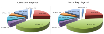
Figure 1: pathophysiological mechanisms of diseases identified as ‘admission’ and ‘secondary’ diagnosis

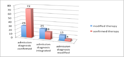
Figure 2: Confirmed/integrated/modified ‘admission’ diagnosis

Discussion and Conclusions

Based on our results, the ‘whole body’ US protocol, as integration of physical exam of the patient admitted in an HDU, might be considered useful for improving diagnostic accuracy and therapeutic management of critically ill patients.
Our US protocol suggested a significant impact on the ‘admission‘ diagnosis. It was ‘integrated’ in 27% of cases and ‘modified’ in 10% of cases. Furthermore the US protocol improved the identification of the pathophysiological and causal mechanisms underlying clinical syndromes and organ failures presented at admission in the HDU. This was true in particular for patients diagnosed with B failure where US evaluation was useful for redirecting the causal diagnosis towards a cardiogenic and/or a pulmonary syndrome.
In patients diagnosed with sepsis, our US protocol was useful for the identification of the septic source and this was useful in order to target the anthibiothic empirical therapy[5].
In conclusion a ‘whole body’ US protocol for evaluation of patients admitted to the HDU from the ED or other units might improve the clinical management allowing to better define the pathophysiological and causal mechanisms underlying presentation syndromes.

Aknowledgments

The research reported in this article was not supported by any fundings . These contents have been presented as a Poster on the last SIMEU National Congress in Naples on the 18th-20th of November 2016.
 

References

  1. A. E. Jones, V. S. Tayal, D. M. Sullivan, J. A. Kline: Randomized, controlled trial of immediate versus delayed goal – directed ultrasound to identify the cause of nontraumatic hypotension in emergency department patients. Crit Care Med 2004 Vol. 32, No 8
  2. SA Haydar, ET Moore, GL Higgins, CB Irish, WB Owens, TD Strout. Effect of bedside ultrasonography on the certainty of physician clinical decisionmaking for septic patients in the emergency department. Ann Emerg Med. 2012 Sep; 60 (3): 346 – 58.
  3. DA Lichtenstein. Point-of-care ultrasound: Infection control in the intensive care unit. Crit Care Med. 2007 May; 35 (5 Suppl): S262 – 7
  4. E. Manno, M. Navarra, L. Faccio, M. Montevallian, L. Bertolaccini, A. Mfochivè, M. Pesce, A. Evangelista. Deep Impact of Ultrasound in the Intensive Care Unit: The ICU – Sound Protocol. Anesthesiology, V 117, No 4, October 2012
  5. A. Leligdowicz, P. M. Dodek, M. Norena, H. Wong, A. Kumar, A. Kumar: Association between Source of Infection and Hospital Mortality in Patients Who Have Septic Shock. American Journal of Respiratory and Critical Care medicine Vol. 189, No 10, May 2014.