Cervicofacial emphysema after a dental hygiene procedure: a case report

Silvia Paiardi1, Sara Baggio2, Carlo Andrea Pivato2, Carlo Fedeli3, Antonio Voza3
  1. Emergency Department, Humanitas Research Hospital, Rozzano (Milano), Italy
  2. Cardiovascular Department, Humanitas Research Hospital, Rozzano (Milano), Italy
  3. Emergency Department, Humanitas Research Hospital, Rozzano (Milano), Italy

Abstract

Subcutaneous emphysema is a rare complication of intraoral surgery; few cases have been reported after minor dental procedures. It usually develops as a sudden painful swelling with crepitus on palpation. It could be misdiagnosed as an allergic reaction or acute post-operative swelling, leading to serious complications, including mediastinitis.
We present a rare case of subcutaneous emphysema extending from the left scalp to the neck, after a dental hygiene procedure. 
The purpose of this report was to provide a brief overview of the literature, report on the case, and review the management of subcutaneous emphysema in the Emergency Department.

Keywords

subcutaneous emphysema, dental procedure, dentistry, air polishing, tooth cleaning 

Introduction

Subcutaneous emphysema is the penetration of gasses into the subcutaneous tissues, leading to a swelling of the affected area. Causes include injury, head and neck surgery, mechanical ventilation, and invasive procedures. A few cases have developed during dental procedures, principally third molar extraction [1] [2], and due to increased mouth air pressure while playing a wind instrument [3] or blowing up a balloon [4]. Emphysema developing after dental procedures is usually limited to the head and neck, with only a few cases involving the mediastinum [5] [6].

We present a rare case of subcutaneous emphysema extending from the left scalp to the neck, following a maxillary dental hygiene procedure.

Case Report

A 40-year-old woman was referred by her dentist to the Emergency Department (ED) of Humanitas Research Hospital, Italy,  because she suddenly developed a large painful swelling from the left scalp to the neck, involving the left eye, during a routine dental hygiene procedure in the upper left molars. Pain was not aggravated by palpation, but subcutaneous crepitus was evident.
At admission, her vital signs were normal, and she did not complain of dysphagia, dyslalia, dyspnea or chest pain. An intraoral examination revealed no visible incision. The airway was not obstructed. Her past medical history was unremarkable. 
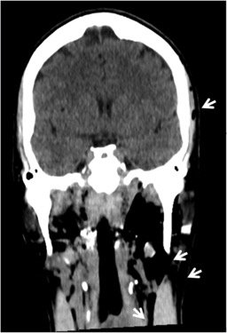
In the suspect of subcutaneous emphysema, she underwent a computed tomography (CT) scan of the maxillofacial region, to examine the extension of the condition; in the absence of chest pain or other signs suggesting the involvement of the mediastinum, chest was not included in the scans. Images revealed significant emphysema of both superficial and deep subcutaneous soft tissues of the left face reaching cranially the left periorbital and temporal region, extending from the masticatory, parapharyngeal and laterocervical spaces until the retropharyngeal space at the height of oropharynx (Figure 1); the emphysema partly continued in the laterocervical soft tissues below the field of view included in the exam. CT scan did not recognize bone lesions. 
In order to exclude involvement of the upper airways, the patient was evaluated by an otorhinolaryngologist; the fibrolaryngoscopy did not find abnormalities in the upper respiratory and gastrointestinal tracts.
Since the patient was asymptomatic, she was discharged with a prophylactic antibiotic therapy (Azithromycin 500 mg daily for 8 days), in order to prevent soft-tissues infection due to the microbial flora from the oral cavity, and a planned short-term follow-up, since the swelling usually resolves spontaneously in few days. 

Discussion

A variety of conditions can cause subcutaneous emphysema, including injury, head and neck surgery, mechanical ventilation, and invasive procedures on the respiratory tract. 
Few cases have been observed during dental procedures, principally third molar extraction [1] [2].Turnbull, in 1900, was the first to describe subcutaneous emphysema and pneumomediastinum developing after dental treatment, when a musician blew a bugle immediately after tooth extraction [3].From 1960 to 2008 a total of 122 cases of emphysema developing after dental treatment were reviewed [5] [7]; some cases have been associated with the use of a dental lasers and with restorative or periodontal treatments using peroxide hydrogen or sodium hypochlorite irrigants, without mucosal incision [5].
In our report, subcutaneous emphysema developed after a simple hygiene procedure; the main risk factors could have been the use of high speed air and sodium hypochlorite (NaOCl)irrigants. Moreover the patient frequently underwent dental hygiene procedures (every four months) and all these three conditions could have lead to the weakening of the periodontal ligament, facilitating the penetration of air into the subcutaneous tissues and the dissection along the planes of the fascial spaces. 
Tubergen et al. reported a similar case after the application of air through intact mucosa, directed into the gingival sulcus to dry teeth [8]. Alonso et al. reported three cases where tooth cleaning using air polishing combined with an abrasive powder was the main risk factor for subcutaneous emphysema, especially when the powder and device were from different manufacturers [9]. Moreover, NaOCl is a commonly used irrigating solution because of its efficacy, but it can also negatively affect the periapical tissues due to its proteolytic action, alkalinity and hypertonicity [10].
The diagnosis of subcutaneous emphysema is mostly clinical based on local signs and symptoms, usually benign and self-limiting: sudden onset of swelling, dysphagia, and crepitus on palpation [6]. Features suggestive of pneumomediastinum are dyspnoea with a brassy voice, chest or back pain, or the Hamman sign – that is the presence of crunching sound with each heartbeat, and nonspecific electrocardiographic changes in the ST-T interval (inverted T waves, ST segment elevation, and shift in the electrical axis) [11]. CT is the most useful imaging technique. 
The differential diagnosis includes allergic reactions, hematomas, cellulites, and angioedema [12]. Trismus and intraoral swelling are typical features of injection hematoma. When the diagnosis is difficult, the best option is empirical treatment as for an anaphylactic reaction until definitive diagnosis. 
Complications include the need for tracheostomy or thoracic drainage, mediastinitis, air embolism, pneumopericardium, pneumothorax, cardiac tamponade, cardiac failure, necrotizing fasciitis [13], and death [14]. 
Although infection is not usually observed in subcutaneous emphysema, this condition may develop due to introduction into the soft tissues of air, microorganisms from the oral cavity, and not sterile water, which may contain Legionella and Pseudomonas subspecies, rendering antibiotic therapy and microbiological monitoring even more critical [6]. The use of a prophylactic antibiotic therapy is recommended and amoxicillin or its equivalent are appropriate

Conclusions

Our case is unusual in terms of the extent of emphysema noted and the simple dental procedure that triggered the problem. We present this case to emphasize the need of a rapid recognition of this condition in the ED, even if the patient has undergone simple endodontic procedure, in order to plan an appropriate management and to avoid rare but serious complications.

Figure 1 Coronal CT image depicting extensive emphysema (arrows) of both superficial and deep subcutaneous soft tissues of the left temporal region, extending from the masticatory, parapharyngeal and laterocervical spaces until the retropharyngeal space at the height of oropharynx.

Acknowledgements

Authors did not receive any financial grants nor other funding. The authors declare that there are no conflicts of interest regarding this paper.

Abbreviations

ED: Emergency Department
CT: Computer Tomography 


References

  1. Yang, T. Chiu, T. Lin, and H. Chan, Subcutaneous emphysema and pneumomediastinum secondary to dental extraction: a case report and literature review,” The Kaohsiung Journal of Medical Sciences, vol. 22, no. 12, pp. 641–645, 2006.
  2. S. McKenzie and M. Rosenberg, “Iatrogenic subcutaneous emphysema of dental and surgical origin: a literature review,” Journal of Oral and Maxillofacial Surgery, vol. 67, no. 6, pp. 1265–1268, 2009.
  3. Turnbull, “A remarkable coincidence in dental surgery,” British Medical Journal, vol. 1, no. 2053, p. 1131, 1900. 
  4. G. Maxwell, K. M. Thompson, and M. S. Hedges, “Airway compromise after dental extraction,” Journal of Emergency Medicine, vol. 41, no. 2, pp. e39–e41, 2011.
  5. Arai, T. Aoki, H. Yamazaki, Y. Ota, and A. Kaneko, “Pneumomediastinum and subcutaneous emphysema after dental extraction detected incidentally by regular medical checkup: a case report,” Oral Surgery, Oral Medicine, Oral Pathology, Oral Radiology and Endodontology, vol. 107, no. 4, pp. e33–e38, 2009. 
  6. Bocchialini G., Ambrosi S., Castellani A., Massive Cervicothoracic Subcutaneous Emphysema and Pneumomediastinum Developing during a Dental Hygiene Procedure, Case Reports in Dentistry Volume 2017, Article ID 7016467, https://doi.org/10.1155/2017/7016467
  7. N. Heyman and I. Babayof, “Emphysematous complications in dentistry, 1960–1993: an illustrative case and review of the literature,” Quintessence International, vol. 26, no. 8, pp. 535–543, 1995
  8. A. Van Tubergen, D. Tindle, G.M. Fox, Sudden Onset of Subcutaneous Air Emphysema After the Application of Air to a Maxillary Premolar Located in a Nonsurgical Field, Operative Dentistry, 2017, 42-5, E134-E138
  9. Alonso, et al. Subcutaneous emphysema related to air-powder tooth polishing: a report of three cases, Australian Dental Journal 2017; 62: 510–515
  10. Tenore, et al. Subcutaneous emphysema during root canal therapy: endodontic accident by sodium hypoclorite, Annali di Stomatologia 2017;VIII(3):117-122
  11. J. Boggess, J. Ronan, N. Panchal, Orbital, Mediastinal and Cervicofacial Subcutaneous Emphysema after Dental Rehabilitation in a Pediatric Patient, Pediatr Dent 2017;39(7):465-7
  12. A. Aslaner, G. N. Kasap, C. Demir, M. Akkas¸, and N. M. Aksu, “Occurrence of pneumomediastinum due to dental procedures,” American Journal of Emergency Medicine, vol. 33, no. 1, pp. 125.e1–125.e3, 2015
  13. C. Karras and J. J. Sexton, Cervicofacial and mediastinal emphysema as the result of a dental procedure,” Journal of Emergency Medicine, vol. 14, no. 1, pp. 9–13, 1996.
  14. B. Aragon, M. Franklin Dolwick, and S. Buckley, “Pneumomediastinumand subcutaneous cervical emphysema during third molar extraction under general anesthesia,” Journal ofOral and Maxillofacial Surgery, vol. 44, no. 2, pp. 141–144, 1986.